Tissues in the Musculoskeletal System
Bone Tissues
There are two types of bone tissue: compact and spongy. The names imply that the two types differ in density, or how tightly the tissue is packed together. There are three types of cells that contribute to bone homeostasis. Osteoblasts are bone-forming cell, osteoclasts resorb or break down bone, and osteocytes are mature bone cells. An equilibrium between osteoblasts and osteoclasts maintains bone tissue.
Compact Bone
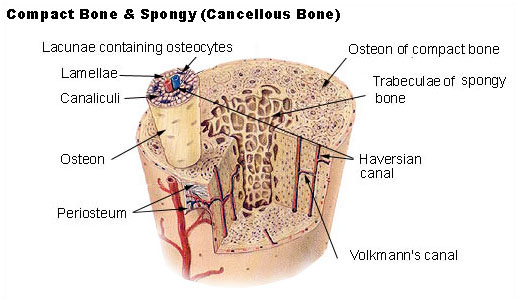
Compact bone consists of closely packed osteons or haversian systems. The osteon consists of a central canal called the osteonic (haversian) canal, which is surrounded by concentric rings (lamellae) of matrix. Between the rings of matrix, the bone cells (osteocytes) are located in spaces called lacunae. Small channels (canaliculi) radiate from the lacunae to the osteonic (haversian) canal to provide passageways through the hard matrix. In compact bone, the haversian systems are packed tightly together to form what appears to be a solid mass. The osteonic canals contain blood vessels that are parallel to the long axis of the bone. These blood vessels interconnect, by way of perforating canals, with vessels on the surface of the bone.
Spongy Bone
Spongy bone is lighter and less dense than compact bone. Spongy bone consists of plates (trabeculae) and bars of bone adjacent to small, irregular cavities that contain red bone marrow. The canaliculi connect to the adjacent cavities, instead of a central haversian canal, to receive their blood supply. It may appear that the trabeculae are arranged in a haphazard manner, but they are organized to provide maximum strength similar to braces that are used to support a building. The trabeculae of spongy bone follow the lines of stress and can realign if the direction of stress changes.
Compact Bone
Compact bone consists of closely packed osteons or haversian systems. The osteon consists of a central canal called the osteonic (haversian) canal, which is surrounded by concentric rings (lamellae) of matrix. Between the rings of matrix, the bone cells (osteocytes) are located in spaces called lacunae. Small channels (canaliculi) radiate from the lacunae to the osteonic (haversian) canal to provide passageways through the hard matrix. In compact bone, the haversian systems are packed tightly together to form what appears to be a solid mass. The osteonic canals contain blood vessels that are parallel to the long axis of the bone. These blood vessels interconnect, by way of perforating canals, with vessels on the surface of the bone.
Spongy Bone
Spongy bone is lighter and less dense than compact bone. Spongy bone consists of plates (trabeculae) and bars of bone adjacent to small, irregular cavities that contain red bone marrow. The canaliculi connect to the adjacent cavities, instead of a central haversian canal, to receive their blood supply. It may appear that the trabeculae are arranged in a haphazard manner, but they are organized to provide maximum strength similar to braces that are used to support a building. The trabeculae of spongy bone follow the lines of stress and can realign if the direction of stress changes.
Muscle Tissue
Muscle tissue is categorized on the basis of a functional property: the ability of its cells to contract. In muscle tissue, the bulk of the cytoplasmic volume consists of the contractile protein fibrils actin and myosin. Muscle is responsible for movement of the body and changes in the size and shape of internal organs. Muscle cells are generally referred to as muscle fibres. Muscle fibres are typically arranged in parallel arrays, allowing them to work together effectively. Our human body contains three types of muscle tissues.
Skeletal Muscle
Skeletal muscle is the most abundant tissue in the vertebrate body. These muscles are attached to and bring about the movement of the various bones of the skeleton.
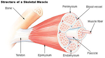
The whole muscle, such as the biceps, is enclosed in a sheath of connective tissue, the epimysium. This sheath folds inwards into the substance of the muscle to surround a large number of smaller bundles, the fasciculi. These fasciculi consist of still smaller bundles of elongated, cylindrical muscle cells, the fibres. Each fibre is a syncytium. The nuclei are oval in shaped and are found at the periphery of the cell, just beneath the thin, elastic membrane. The sarcoplasm also have many alternating light and dark bands, giving the fibre a striped or striated appearance. Each muscle fibre is made up of many smaller units, the myofibrils. Each myofibril consists of small protein filaments, known as actin and myosin filaments.
Smooth Muscle
Smooth muscle tissue is made up of thin-elongated muscle cells, fibres. These fibres are pointed at their ends and each has a single, large, oval nucleus. Each cell is filled with a specialised cytoplasm, the sarcoplasm and is surrounded by a thin cell membrane, the sarcolemma. Each cell has many myofibrils which lie parallel to one another in the direction of the long axis of the cell. They are not arranged in a definite striped (striated) pattern, as in skeletal muscles. Smooth muscle fibres interlace to form sheets or layers of muscle tissue rather than bundles. Smooth muscle is involuntary tissue, meaning it is not controlled by the brain. Smooth muscle forms the muscle layers in the walls of hollow organs such as the digestive track, the walls of the bladder, the uterus, various ducts of glands and the walls of blood vessels .
Cardiac Muscle
This is a unique tissue found only in the walls of the heart. Cardiac Muscle Tissue shows some of the characteristics of smooth muscle and some of skeletal muscle tissue. Its fibres , like those of skeletal muscle, have cross-striations and contain numerous nuclei. However, like smooth muscle tissue, it is involuntary. Cardiac muscle differ from striated muscle in the following aspects: they are shorter, the striations are not so obvious, the sarcolemma is thinner and not clearly discernible, there is only one nucleus present in the centre of each cardiac fibre and adjacent fibres branch but are linked to each other by so-called muscle bridges. The spaces between different fibres are filled with areolar connective tissue which contains blood capillaries to supply the tissue with the oxygen and nutrients.
Skeletal Muscle
Skeletal muscle is the most abundant tissue in the vertebrate body. These muscles are attached to and bring about the movement of the various bones of the skeleton.
The whole muscle, such as the biceps, is enclosed in a sheath of connective tissue, the epimysium. This sheath folds inwards into the substance of the muscle to surround a large number of smaller bundles, the fasciculi. These fasciculi consist of still smaller bundles of elongated, cylindrical muscle cells, the fibres. Each fibre is a syncytium. The nuclei are oval in shaped and are found at the periphery of the cell, just beneath the thin, elastic membrane. The sarcoplasm also have many alternating light and dark bands, giving the fibre a striped or striated appearance. Each muscle fibre is made up of many smaller units, the myofibrils. Each myofibril consists of small protein filaments, known as actin and myosin filaments.
Smooth Muscle
Smooth muscle tissue is made up of thin-elongated muscle cells, fibres. These fibres are pointed at their ends and each has a single, large, oval nucleus. Each cell is filled with a specialised cytoplasm, the sarcoplasm and is surrounded by a thin cell membrane, the sarcolemma. Each cell has many myofibrils which lie parallel to one another in the direction of the long axis of the cell. They are not arranged in a definite striped (striated) pattern, as in skeletal muscles. Smooth muscle fibres interlace to form sheets or layers of muscle tissue rather than bundles. Smooth muscle is involuntary tissue, meaning it is not controlled by the brain. Smooth muscle forms the muscle layers in the walls of hollow organs such as the digestive track, the walls of the bladder, the uterus, various ducts of glands and the walls of blood vessels .
Cardiac Muscle
This is a unique tissue found only in the walls of the heart. Cardiac Muscle Tissue shows some of the characteristics of smooth muscle and some of skeletal muscle tissue. Its fibres , like those of skeletal muscle, have cross-striations and contain numerous nuclei. However, like smooth muscle tissue, it is involuntary. Cardiac muscle differ from striated muscle in the following aspects: they are shorter, the striations are not so obvious, the sarcolemma is thinner and not clearly discernible, there is only one nucleus present in the centre of each cardiac fibre and adjacent fibres branch but are linked to each other by so-called muscle bridges. The spaces between different fibres are filled with areolar connective tissue which contains blood capillaries to supply the tissue with the oxygen and nutrients.
